Professors: Ana Paula Moreira Barboza / Mariana de Castro Prado
Research Lines:
- Characterization of two-dimensional materials using Scanning Probe Microscopy.
- Characterization of cancerous cells and tissues using Scanning Probe Microscopy.
SPM (Scanning Probe Microscopy) is the generic name for a family of microscopy techniques that use a mechanical probe to detect various physical quantities to study surface properties. Each technique arises from the type of probe/sample interaction being monitored, allowing not only high-resolution morphological analysis of the samples but also providing information on other intrinsic properties such as hardness, viscoelasticity, and magnetic and electrical properties. It is also possible to manipulate samples physically or chemically using the SPM probe as a modification tool.
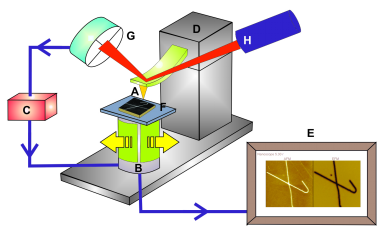
Despite providing different types of information, all SPM techniques are based on the same operating principle. A mechanical probe is placed in contact or very close to the sample surface, creating a highly localized interaction. A piezoelectric scanner moves the sample laterally relative to the probe, following a scanning pattern. A monitoring mechanism detects the variation in the probe/sample interaction during the scan through the laser position change on a photodetector, and this information is passed to a feedback system that controls the probe's vertical position. A computer controls the process, moving the scanner, receiving, and converting the data to form the sample image.
Schematic drawing of the common components of all Scanning Probe Microscopes.

Schematic drawing of the common components of all Scanning Probe Microscopes. (A) mechanical probe; (B) piezoelectric scanner; (C) probe/sample interaction monitoring mechanism; (D) preliminary probe positioning mechanism over the sample; (E) computer controlling the entire system; (F) sample.
Two main research lines are currently being developed:
-
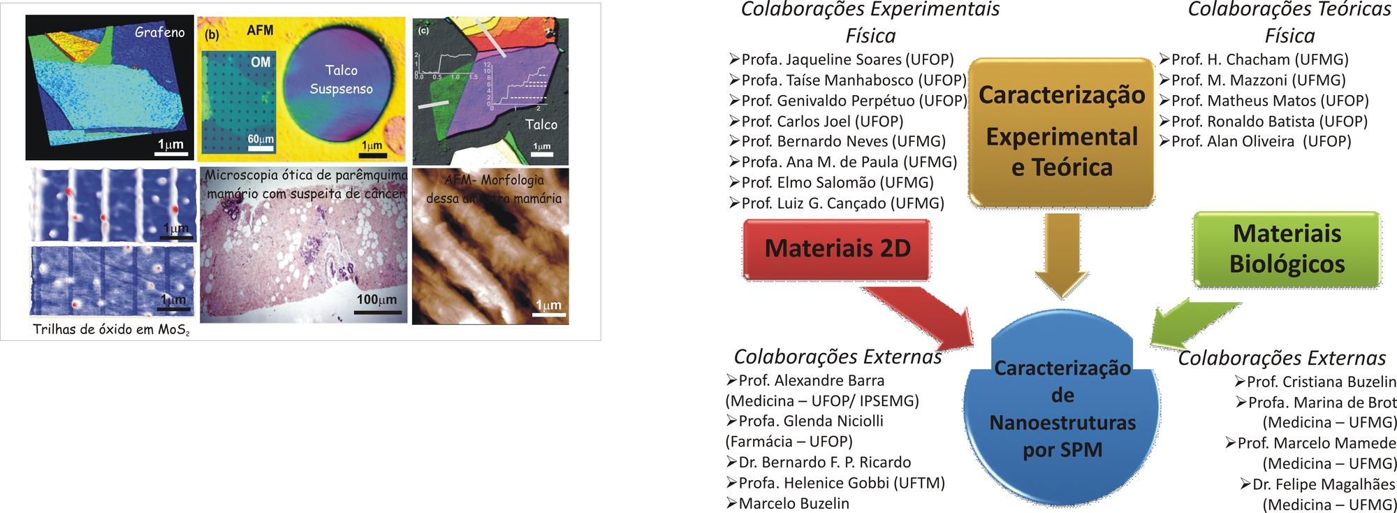
Characterization of two-dimensional materials using Scanning Probe Microscopy: In this project, we study the electromechanical and magnetic properties of exfoliable two-dimensional materials such as graphene, molybdenum disulfide, boron nitride, and talc. The goal is to analyze new properties/materials to optimize their use in electronic devices. This project is developed in collaboration with two major theoretical groups: the Laboratory of Electronic Structure at UFMG and the Laboratory of Computational Simulations at UFOP.
-
Characterization of cancerous cells and tissues using Scanning Probe Microscopy: The Scanning Probe Microscopy technique stands out for its great potential in studying the mechanical properties of different biological materials. In addition to allowing a morphological analysis of the surface, it is possible to perform measurements in regions in a highly localized and controlled manner. The goal of this project is to demonstrate that the Scanning Probe Microscope can be used as an additional tool in cancer diagnosis. The proposal involves using the probe to perform comparative tests of mechanical properties in healthy and diseased tissues. This is an interdisciplinary project that unites the Departments of Physics and Medicine at both UFOP and UFMG in a novel initiative at both institutions. We have already gathered all the health professionals responsible for the collection and pathological analysis of the material for subsequent comparison with SPM measurements. Two types of cancer will be analyzed: breast cancer and prostate cancer. Regarding cells, we will study bladder tumor cells.
All the infrastructure (sample preparation rooms, equipment setup) necessary for the development of the proposed projects is being set up in the Department of Physics at UFOP, and until it is completed, experiments are being conducted in collaboration with the Department of Physics at UFMG, where there are three operational SPM devices. It is important to highlight that this interaction will allow students to learn and practice the SPM technique, which is fundamental to the development of the projects, as well as give them greater exposure to procedures and equipment applied to Nanoscience and Nanotechnology.
Below are some images associated with the previously mentioned projects and established collaborations:

